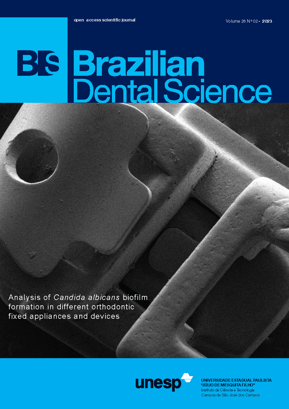
Analysis of biofilm formation by Candida albicans in different types of orthodontic fixed appliances and devices
DOI:
https://doi.org/10.4322/bds.2023.e3440Abstract
Objective: in this study, biofilm formation by Candida albicans in fixed orthodontic appliances was evaluated. Material and Methods: a total of 300 conventional metal brackets (MC), ceramic (CB), self-ligation (SLB), nickel-titanium (NiTi), and nickel-chromium (NiCr) wires, and ligatures types were organized into thirty groups (n=10). To induce biofilm formation, brackets, wires, and ligatures were joined, sterilized, placed in 24-well plates, contaminated with standardized suspensions of C. albicans (107 cells/mL), and incubated at 37 °C for 48 h with shaking. The biofilms formed were detached using an ultrasonic homogenizer, and suspensions were serially diluted and plated on Sabouraud dextrose agar to determine colony-forming units per mL. Scanning electron microscopy was performed before and after the biofilm formation. Results: lower amount of biofilm formation was observed in the MC group than in the CB and SLB groups (p<0.0001). SLB and CB showed similar biofilm formation rates (p=0.855). In general, the cross-sectional wires .018”x.025” showed higher biofilm formation when associated with the three types of brackets. When brackets, wires, and ligatures were associated, the sets with NiCr wires and SSL ligatures with MC brackets (p=0.0008) and CB (p=0.0003) showed higher biofilm formation. Conclusion: thus, brackets of MC with NiTi and NiCr wires showed lower biofilm formation, regardless of the ligature and cross-sectional or gauge of the wire and, MC and CB brackets with NiCr wires and SSL ligatures were more likely to accumulate biofilms.
KEYWORDS
Biofilms, Candida albicans, Orthodontic appliances, Orthodontic brackets, Scanning electron microscopy.
Downloads
Published
How to Cite
Issue
Section
License
Brazilian Dental Science uses the Creative Commons (CC-BY 4.0) license, thus preserving the integrity of articles in an open access environment. The journal allows the author to retain publishing rights without restrictions.
=================


